Acoustic Shadow Moiré A Solution for 3D Biomedical Imaging - Juniper Publishers
Juniper Publishers - Open Access Journal of Engineering Technology
Abstract
This article introduces a new 3D imaging technique
for biomedical applications. The new technique uses shadow Moiré
principles to produce 3D acoustic imaging. Optical shadow Moiré
principles have been used in many industrial applications, but the
acoustic counterpart has not been seriously investigated. The
feasibility of using acoustic shadow Moiré in 3D imaging have been
investigated by the author's research group. This review argues that
using acoustic shadow Moiré mechanism can provide surface and volumetric
3D biomedical imaging technology with the safety and resolution needed
for medical radiology.
Keywords: : Shadow Moiré; Acoustic shadow Moiré; 3D imaging; Acoustic imaging; Medical diagnosisIntroduction
The Moiré effect is a result of the interference
between two repetitive structures or images; e.g. grids, gratings, and
their shadows. The interference between the superimposed patterns
creates an image of alternating dark and bright areas. The shape of this
image is sensitive to the topology of the surface on which the waves
are reflected.
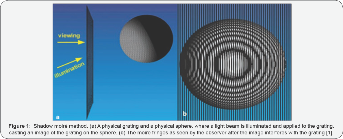
An example of Moiré effect is shown in Figure 1 [1].
In part (a) of the figure, the lower arrow represents the illumination
ray. The upper arrow represents the viewing camera or eye. The
illuminating wave goes through the physical grating creating an image of
the grating on the sphere. The reflected image of the grating from the
sphere surface goes again through the physical grating to the observer's
eye. The observer sees the image shown in part (b). As can be seen from
the figure, the image that the observer sees is a sphere, formed as a
result of patterns interference. The intensity and the shapes of the
pattern determine the vertical dimension, which is the curvature of the
sphere. If the imaged object was a plate, the pattern would show the
topology of its surface. In other words, Moiré pattern gives a 3D image
of the surface.

The resolution of the image is determined by the
grating pitch and the beam wavelength. A smaller pitch can be used with a
shorter wavelength to provide higher resolutions or sharper images.
However, to create the real image of the object and show the contour of
the surface topology, signal processing algorithms are used. For optical
shadow Moiré imaging, many advanced algorithms have been developed and
are commercially available.
Optical shadow Moiré has been used in many
applications. It has been used in contour mapping of the human body to
detect any asymmetries caused by certain illness, such as scoliosis
deformation in the body. This technique can also be used in strain
measurements and in studies of metal deformation. The size of the
strain, the deformation, or the buckling can be seen in the shadow Moiré
photographs. Shadow Moiré can also be used in manufacturing. For
example, it can be used to establish the contours of sheet metal
stampings. It can also be used for quality- control, by comparing the
contour of a manufactured object with that of a master object. Shadow
Moiré has recently been adopted by the electronics industry to measure
the war page or the co planarity of microelectronic devices and
integrated circuits.
Acoustic Shadow Moiré
Can shadow Moiré fringes be formed, if acoustic waves
are applied? This question motivated the author to investigate the
feasibility and practicality of using acoustic shadow Moiré imaging.
This issue was investigated experimentally and using numerical analysis.
Ultrasound wave with a MHz frequency was applied to an aluminum grating
and reflected from a flat surface to interfere with the grating again
before being detected by an ultrasound camera [2].
The image from the camera showed that interference patterns were
formed, providing the proof that Moiré fringes can be created using
ultrasound waves. Acoustic Moiré fringes were also investigated using
finite element numerical analysis [3]. COMSOLTM, a multi physics numerical analysis program, was used in this study [4].
The results from the experimental and numerical studies were found to
be in full agreement, proving the existence of acoustic shadow Moiré
effect.
Discussion and Conclusion
In diagnostic radiology, ultrasound is preferred
because it has two advantages: (a) it does not pause lasting harm to the
living cells, and (b) it can penetrate tissues with a penetrating power
related to its frequency. Waves with lower frequencies penetrate deeper
that those with high frequencies. Therefore, for deep abdomen imaging,
2.5MHz is used; for gynaecological imaging, 3.5MHz is used; for vascular
imaging, 5.0MHz is used; for breast and thyroid imaging, 7.5MHZ is
used; for superficial veins and masses imaging, 10.0MHz is used; and for
musculoskeletal imaging, 15MHz is used. In conventional ultrasound
imaging, a beam is applied to the tissue and the scattered beam is
collected to form an image. The diagnosis is made based on the
inspection and interpretation of the different intensities of the
reflected beams. Ultrasound imaging is good for initial qualitative
check, but for a more conclusive diagnosis, high resolution techniques,
such as X-ray or MRI, are normally used.
Acoustic shadow Moiré imaging is expected to result
3D imaging with sub millimeter resolutions. This is because the
resolution (size of minimum feature) is proportional to the wave length.
For a 2.5 MHz wave, a resolution of sub millimeter can be obtained. The
resolution will increase for higher frequencies. This technique can
also be holographic and multi-shaded volumetric imaging. The author
expects that by using different frequencies, not only the surface of the
object can be imaged, but the volume from inside can also be visible.
This would be possible by using beams with different frequencies to
image at different penetrations. This technique can also be used to
produce videos to capture real time change in the body.
Ultrasound shadow Moiré imaging still needs a great
deal of development. In this regards, practical imaging systems need to
be developed and tested. Sophisticated digital signal processing
algorithms, especially for real time imaging, must be developed before
the full potential of this technique can be realized.
Conclusion
In conclusion, the research conducted by the author
and his group has proven, experimentally and theoretically, that shadow
Moiré effect can be created using acoustic waves. With the advantage of
ultrasound in biomedical imaging, shadow Moiré can provide safe 3D
imaging with high resolution. The images can be holographic as well as
volumetric, where the surface as well as the interior can be imaged.
This simple imaging technique is expected to move biomedical imaging to
new frontiers, where using different frequencies and different grating
pitches can provide more degrees of freedom.
For more articles in Open Access Journal of
Engineering Technology please click on:
https://juniperpublishers.com/etoaj/index.php


Comments
Post a Comment